 Home:
Meeting
Highlights: Posters

Reduction
Mammoplasty to Improve Breast Conservation Therapy Results in Patients
with Macromastia
Newman, Lisa A; Kuerer, Henry Mark; Gunter, Joseph; Ames, Fred C;
Ross, Merick I; McNeese, Marsha D; Robb, Geoffery; Singletary, S
Eva
BACKGROUND:
Macromastia
has been considered a contraindication to breast conservation therapy
(BCT) because of difficulties with radiation therapy. This study
evaluates the feasibility of bilateral reduction mammoplasty as
a component of BCT for breast cancer patients with pendulous breasts.
METHODS:
Of 153 patients
undergoing reduction mammoplasty at the University of Texas M.D.
Anderson Cancer Center, 28 were identified as breast patients with
macromastia receiving BCT. Median follow-up was 23.8 months.
RESULTS:
Median patient
age was 55 years. Nearly all patients were described as obese. Median
weight of the reduction mammoplasty specimen on the cancerous side
was 766 grams. One patient (4%) required completion mastectomy for
inadequate margin control. Major postoperative complications occurred
in two patients (7%). There were no major post-radiation complications.
Patient survey revealed a satisfaction rate of 86%.
CONCLUSION:
Bilateral reduction
mammoplasty is a reasonable and safe option for breast cancer patients
with macromastia who desire BCT.
Breast conservation
therapy, consisting of a margin-negative segmental mastectomy and
breast irradiation, is a standard and oncologically safe treatment
modality for patients with early stage breast cancer, and for many
patients with locally advanced disease, if their tumors can be downstaged
sufficiently with induction chemotherapy. Multicentric lesions,
or a medical contraindication to chest wall irradiation, however
mandate mastectomy for definitive locoregional disease control.
The presence of obesity or other body habitus associated with large,
pendulous breasts can complicate the efficacy and suitability of
both treatment approaches.
A unilateral
mastectomy for a woman with macromastia can cause an unacceptable
degree of asymmetry that will make prosthesis fitting very difficult,
and results in substantial imbalance that can adversely affect quality
of life. On the other hand, delivery of radiotherapy to a bulky
and ptotic breast can be very challenging technically, and may result
in excessive radiation toxicity.
Bilateral reduction
mammoplasty in conjunction with tumor-directed segmental mastectomy
is a surgical technique that can potentially improve the efficacy
of radiation therapy in this setting, alleviate the neuropathic
symptoms that can accompany macromastia, and increase rates of breast-preserving
surgery for breast cancer patients. However, this option is frequently
overlooked by surgeons managing newly-diagnosed breast cancer patients,
and consequently there is limited reported data regarding the success
rate of this approach. The purpose of this study was to evaluate
the outcome of reduction mammoplasty in conjunction with breast
conservation therapy as measured by complication rates and patient
satisfaction.
The medical
records were reviewed for 153 patients coded as having undergone
reduction mammoplasty at the University of Texas M.D. Anderson Cancer
Center between 1994 and 1999. Twenty-eight patients were identified
as breast cancer patients who underwent bilateral reduction mammoplasty
as a component of conservation therapy because of macromastia or
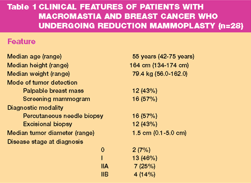
excessively ptotic breasts (Table I). The mammoplasty incision followed
the general pattern described by Wise. Attempts were made to contact
all patients via telephone for participation in a patient satisfaction
survey. All contacted patients were queried regarding the need for
any additional revisional breast plastic reconstruction surgery.
Patient satisfaction with result was scored as excellent, modestly
satisfied or unsatisfied (patient regretted undergoing the reduction
mammoplasty). Median follow-up from date of surgery was 23.8 months.

The discussion
regarding reduction mammoplasty was initiated by the patient's request
for a smaller breast size in 11 cases (39%) and by the physician
because of suspected radiation-related difficulties in the remaining
17 cases (61%). Three patients received neodajuvant chemotherapy
for locally advanced primary tumors (greater than 4 centimeters).
Median weight
of the reduction mammoplasty specimen (including the weight of the
tumor –bearing segmental mastectomy tissue) on the cancerous side
was 766 grams (range, 150-3,250 grams) and 645 grams on the non-cancerous
side (range, 150-3,230 grams). Adequate margin control (more than
2 mm microscopically negative margin) was achieved in all except
for two patients (7%). Extensive ductal carcinoma in situ was found
in one patient, necessitating completion mastectomy; a single microscopic
focus of cancer was found at the deep margin, approaching the pectoral
fascia in the other patient. No significant pathology (atypia, in
situ, or invasive cancer) was found in any of the contralateral
reduction mammoplasty specimens.
An axillary
lymph node dissection was performed via a separate axillary incision
in 26 patients (93%) with a median of 15 lymph nodes identified
(range, 6-33). Six patients underwent lymphatic mapping and sentinel
lymph node dissection; a sentinel lymph node was identified in four
of these cases. In one patient with a failed lymphatic mapping procedure
preoperative chemotherapy had been administered , and in the other
patient a prior excisional biopsy of the primary tumor had been
performed. Both of these patients underwent completion axillary
lymph node dissections. In three of the four successful mapping
procedures a completion axillary lymph node dissection was performed,
revealing no false negative cases, and in one of these patients
the sentinel lymph node was the isolated site of axillary metastasis.
There were no
postoperative complications in 18 of the 28 patients (64%). Minor
wound infections requiring oral antibiotics developed in five patients
(18%). Major wound complications developed in two patients (7%):
one experienced significant cellulitis that was treated with a course
of intravenous antibiotics, and the other developed incisional necrosis
requiring surgical debridement.
At the time
of this review, 21 patients have completed their postoperative radiation
therapy, with post-radiation sequelae described as mild erythema
in 11 (53%) and no notable adverse effects in the others.
With a median
follow-up of 23.8 months from the date of surgery, no patients have
experienced a local recurrence, and 2 (7%) distant treatment failures
have been detected. Twenty-seven patients (96%) are alive; 1 (4%)
with stable metastatic disease and 26 (93%) without evidence of
disease.
A telephone
survey regarding outcome and patient satisfaction was completed
by 14 patients. Two (14%) reported having to undergo minor additional
revisional/correctional plastic surgery on their breasts, and 12
(86%) reported being very satisfied with their final cosmetic result.
Only 2 (14%) expressed regret regarding their surgery because of
poor cosmetic outcome and wished they had undergone a mastectomy
instead.
Breast conservation
therapy for early stage breast cancer includes a segmental mastectomy
with an attempt obtain a gross negative margin of at least one centimeter,
is the treatment of choice for patients with unicentric primary
breast tumors under four centimeters in greatest diameter. Postoperative
breast radiation therapy is an essential component of breast conservation
therapy, and results in 12-year local recurrence rates of 10% compared
to 39% seen in women who undergo lumpectomy without irradiation.
For women with locally advanced breast cancers, breast-preserving
therapy remains an option if the tumor can e appropriately downstaged
with induction chemotherapy, and postoperative breast irradiation
yields local recurrence rates that are similar to those seen with
early stage disease. It is clear that the ability to deliver adequate
radiation therapy is critical for successful breast conservation
therapy.
Obesity is a
risk factor for breast cancer development in postmenopausal women
because of the increased rate of conversion of the adrenal estrogen
precursor androstenedione, to estrone by enzymes located in fat
cells. The predominantly fatty replaced breast is generally easier
to screen for mammographic densities suggestive of small cancers,
and a unilateral mastectomy for the very large-breasted woman can
result in an excessively uncomfortable degree of asymmetry and imbalance.
It might therefore be inferred that obese women with large, fatty
breasts should be appropriate, and perhaps better candidates for
breast conserving treatment of early-stage disease.
However, breast
cancer patients with macromastia (whether related to obesity or
individual body habitus) present particular challenges to the radiation
oncologists. The large breast may require higher energy photons
to ensure radiation delivery to the deeper tissue, and the dose
inhomogeneity that can result may lead to significant radiation
toxicity to the skin. The very ptotic breast may also be difficult
for reproducible fixation and positioning during the many treatments,
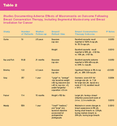
and significantly worse aesthetic results form breast-conserving
treatment associated with large, pendulous breasts have been documented
in several series, as shown in Table II.
This series
demonstrates that bilateral reduction mammoplasty is an excellent
maneuver to improve breast conservation therapy feasibility in women
with macromastia. The safety of this approach is affirmed by the
finding of only two patients experiencing significant postoperative
complications, and no major radiation-related complication developing
and excellent patient satisfaction rate was reported. In addition
, lymphatic mapping with sentinel lymph node biopsy was performed
in only a small number of patients, but our results suggest that
the identification rate and accuracy of the procedure are not significantly
impaired by the reduction mammoplasty.
In summary,
we believe that bilateral reduction mammoplasty can be performed
safely at the time of definitive breast cancer surgery and prior
to breast irradiation in patients with macromastia. Breast conservation
therapy has previously been considered relatively contraindicated
in this patient population. Utilization of this technique should
improve the ability to deliver the radiation component of breast
conservation therapy to women with large, pendulous breast with
acceptably low complication rates.

|
FIGURE
1A
|
FIGURE
2A
|
 |
 |
|
FIGURE
1B
|
FIGURE
2B
|
 |
 |
|
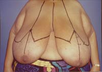
Fig
1.A. Preoperative photograph of breast cancer patient
with macromastia with skin markings for bilateral reduction
mammoplasty.
Fig
1.B. Postoperative photograph of same patient following
resection of over 3000 g of tissue from each breast.
|
Fig
2.A,B. Pre- and Post-operative photographs (respectively)
of breast cancer patient with macromastia and bilateral reduction
mammoplasty. |
Top
of Page
|


