|
 Home:
Meeting
Highlights: Posters
Home:
Meeting
Highlights: Posters

The
Intradermal Sentinel Node: Update 2000
Owen DH
This poster
updates my experience with sentinel node biopsy utilizing a purely
intradermal injection of radioactive material. Most surgical trials
addressing the accuracy of sentinel node biopsy use an intratumoral
injection of blue dye, radioactive material, or both. However, based
on my 5-year experience using the technique described below, it
is clear that the intradermal route provides identical node positivity
data while obviating the need for image-guided injections into tumor
beds and biopsy cavities. Perhaps more importantly, there is no
need for the “oncologically unpleasant” massage of the
tumor since the radioactive material flows swiftly to the axilla
upon injection, with very high counts.
This poster also addresses the feasibility of sentinel node biopsy
in elderly patients. Here the average age of patients in this study
is 71 years, characteristic of the patient population in Palm Beach
County, Florida. This poster describes a large single-surgeon experience
wherein the sentinel node is identified solely with an intradermal
injection. This series is updated yearly since
1997.
From June 1995
to Nov. 2000, a total of 321 patients have
undergone this procedure. 14 patients were omitted early
in the series (learning curve and probe malfunctions), thus yielding
a series of 307, which comprises this update.
As the technique was described at great length at the SABCS in 1998,
I will only list a few salient points.
1. All patients with operable cancer were included.
2. The injection technique has not changed over the past 5 years:
1 millicurie of filtered tech-99 sulfa colloid
is injected in 3-4 aliquots to raise a
small intradermal wheal, about 1 inch away from the areola
in the same quadrant as the tumor. Using the areola as a clock,
the injections were placed at “5 minute” intervals in
the appropriate quadrant.
3. Pre-op scintigram was obtained.
4. The lumpectomy or mastectomy was performed first. The
tumor bed was not massaged.
5. The axilla was exposed widely after
incision of the clavipectoral fascia.
6. The axilla was then carefully palpated and probed with the Navigator.
7. A sentinel node was defined as a palpable
metastatic node, or a scintigraphically detected node with a count
10 times greater than the background. (Rarely a 5:1 ratio
was obtained).
8. After processing, multiple sections were performed on all sentinel
nodes, with a minimum of three levels, 40 microns apart on each
block. Immunoperoxidase staining for cytokeratins was performed
on all sentinel nodes.
9. All patients underwent surgical treatment and follow up by this
surgeon, with exam and mammogram of the treated breast every 6 months
for lumpectomy patients, and exam every 6 months with contralateral
mammogram every year for mastectomy patients.
Fig 1: This
is an old population base (av.71yrs)
compared to most other series, where the average age tends to be
in the 50’s.
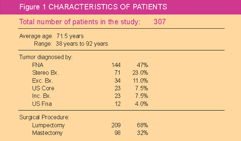
Fig 2: Co-morbidity and loss of follow up was significant with over
8% of patients dying from other disease. In addition close to 18%
of patients have been lost to follow up.
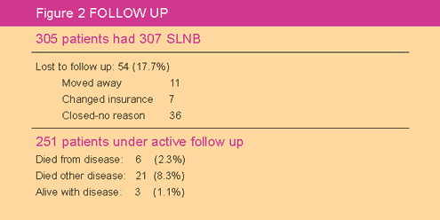
Fig 3,4: Characteristics
of the tumors and locations of the sentinel nodes have remained
unchanged over the years2,3. The average tumor
size has remained 1.5 - 1.6 cm. A sentinel node was found
in every case, either scintigraphic or grossly metastatic. Every
primary sentinel node was found in the level one region of the axilla
in a consistent location with one exception.
The overall rate of SLN positivity for invasive cancer was 31.1%.
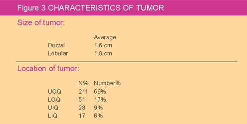
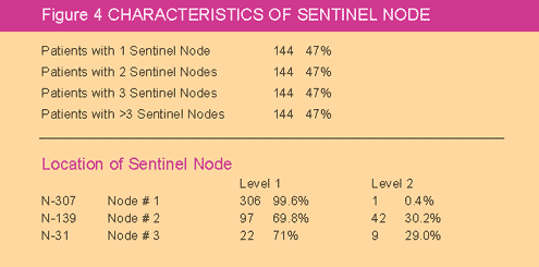
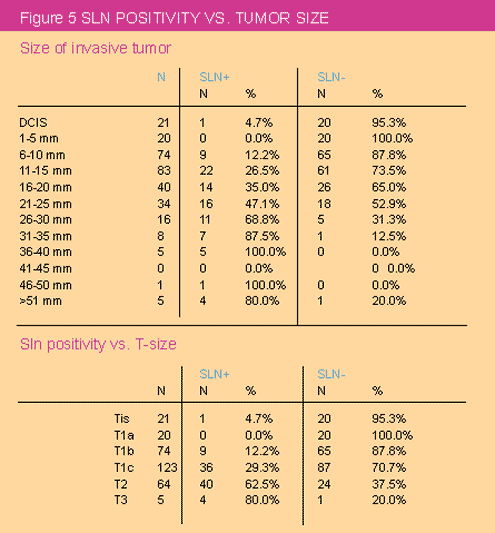
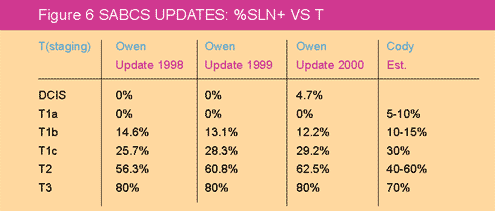
Fig 5,6: The
%-sln positivity vs. T-size has continued to remain constant over
5 years with each update. This falls within the predictive guidelines
suggested by Cody1. The false negative rate
was 2/321 or .6%. There has been one axillary recurrence.
This may have been a “persistence” since it was found
6 months after performing SLNB in an elderly woman where the sentinel
node was grossly positive and a surrounding node was negative. The
patient developed a palpable mass in the axilla. Completion axillary
node dissection revealed no other metastatic nodes and she is alive
and well one year later.
1. The “intradermal” sentinel node is easily and consistently
identified without the need for tumor massaging or imaging.
One or several intradermal injections will result in a rapidly visualized
node found in the level one region of the axilla.
2. In a series of 321 patients, the false negative
rate appears to be dramatically reduced if the axilla is opened widely
and the clavipectoral fascia incised, to allow careful palpation of
the axilla. A negative pre-op scintigram may alert the surgeon to
the possibility of a tumor filled sentinel node near the scintigraphically
positive node.
3. The % SLN positivity for each incremental
increase in T size in this patient population is consistent with that
in other reported series despite different methods of injection2,3.
4. The overall % SLN positivity vs. T size in this elderly population
matches that in other series where average patient age is in the 50’s2,3.
Advanced age is not a barrier to the
successful performance of SLNB.
5. Although this was not specifically addressed in this poster, the
rate of micrometastases in cases of DCIS, T1a and T1b (non-palpable)
continues to provoke controversy. At this time, in our community setting,
we submit cases of H&E negative, immunoperoxidase positive micromets
for second opinion, to “university” pathologists. We specifically
request comment on the biologic significance
of both the small numbers of cells present in the node and their location
within the lymph node itself. In four cases submitted, recently,
all “mets” have been deemed to be biologically insignificant
by virtue of their non-malignant cytological appearance, as well as
by their location in the sinus of the node.
6. Surgical trials underway should strive for uniformity in pathologic
analysis of micrometastatic disease in cases of DCIS and T1a, and
b. Perhaps, using numbers of metastatic cells, their cytological appearance,
and their location in the node, pathologists can develop standards
assessing the biologic significance of
such metastases when they are found. This may ultimately prove to
be more important than the striving for uniformity in injection techniques.
Data such as this attests to the irrelevance
of the injection technique in the determination of biologically significant
sentinel node metastases.
7. As always, no funds were ever used to support this ongoing series…it
continues to be a labor of love. I thank the Selection Committee for
allowing me to update this data here.
Bibliography
1. Cody, Hiram S. et.al, Oncology. January 1999
2. Owen, DH. CD-ROM: SABCS Poster, Dec 1998
3. Owen, DH. CD-ROM: SABCS Poster, Dec 1999
Top of Page
|
