 Home:
Meeting
Highlights: Posters
Home:
Meeting
Highlights: Posters

A Decade of Sentinel Lymph Node Mapping in Breast Cancer
A
Decade of Sentinel Lymph Node Mapping in Breast Cancer:
A Hypothesis - Driven Journey Toward a New Paradign
Grube BJ, Hansen NM, Turner RR, Brennan MB, Krasne DL, Glass EC,
Giuliano AE
Axillary lymph
node dissection (ALND) has been an integral component of the surgical
management of invasive breast carcinoma. For much of the 20th century,
breast cancer was believed to spread in an orderly process. Radical
en bloc resection of the breast, adjacent tissues and its draining
lymphatics was the standard of surgical therapy. During the 1970's,
this concept was challenged by Fisher, who suggested that breast
cancer was a systemic disease from inception and that the surgical
management of this disease should be reevaluated. Out of this challenge
grew the trials that confirmed breast conservation treatment of
early breast cancer resulted in equivalent long term survival.
During the 1980's,
Morton conducted experiments to determine if intraoperative lymphatic
mapping (ILM) could replace complete lymph node dissection as a
low morbidity procedure for staging of melanoma. In October of 1991,
Giuliano began to test the feasibility of ILM and sentinel lymph
node dissection (SLND) for patients with breast cancer to eliminate
routine ALND without losing the staging accuracy derived from complete
ALND. During the past decade SLND at the JWCI evolved as a technique,
enhanced axillary staging, was validated as a concept, demonstrated
no adverse outcome and decreased morbidity. The challenge for the
next decade is validation of the concept of SLND in multi-institutional
trials, determination of the clinical relevance of SLN assessment
in staging and its role for directing oncologic treatment algorithms
for better comprehensive treatment of breast cancer.
|
1991: The First Challenge Surgical Feasibility |
Can a sentinel
draining lymph node be identified intraoperatively in breast cancer?
In 1991, there was no established protocol for ILM for breast cancer.
The purpose of the pilot study was to establish the methodology
of ILM and SLND in breast cancer and to determine the feasibility,
safety and accuracy of ILM for early stage breast cancer (1).
Between October
1991 and February 1, 1994, 174 patients underwent planned ILM and
SLND followed by a Level I, II and some III ALND. This study was
designed to determine the quantity of isosulfan blue dye (0.5-10
cc), the appropriate site of injection (tumor vs parenchyma vs biopsy
site) and the length of time to allow for lymphatic flow from injection
to axillary incision (1 to 20 minutes) necessary for identification
of putative lymphatics and SLN.
| Figure
1 |
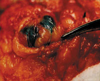 |
This first developmental
study established the guidelines of 3-5 cc isosulfan blue, injected
peritumorally in breast parenchyma or in the wall of the biopsy
cavity and a standard time interval of 5 minutes. Figure 1 demonstrates
the typical appearance of blue lymphatic channel tracking to sentinel
blue nodes (Figure 1).
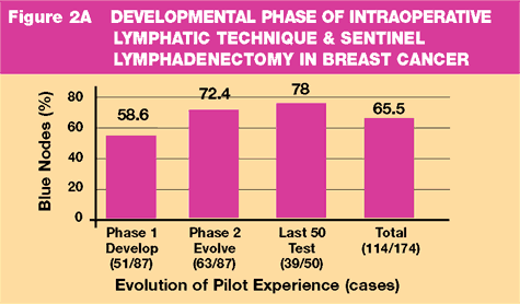
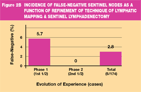
During Phase
1 (Developmental Phase), the ability to identify the SLN was 58.6%
with an accuracy of 94.3%. As the technical variables were refined
and the method evolved (Phase 2), the ability to identify the SLN
improved. The last 50 test cases demonstrated a 78% ability to identify
the SLN with 100% accuracy. Data shown in Figure 2A and B.
- Intraoperative
lymphatic mapping and sentinel node identification in breast cancer
was technically feasible, safe and without added complications
- Injection
of 3-5 cc 1% isosulfan blue followed by surgery ILM in 5 minutes
resulted in identification of the SLN
- SLND identifies
the first ("sentinel") axillary lymph node draining
the primary tumor and most likely node to harbor metastases
- Sentinel
lymph nodes were found in Level I or II, not Level III
- The SLN is
an accurate predictor of the status of the axilla
| Future
Directions Resulting From Pilot Study |
- Further refinements
in the procedure are likely to improve accuracy
- Future role
of ALND may be modified by status of SLN
| The
Second Challenge Histopathologic Evaluation |
Does routine
histologic evaluation of ALN provide adequate information of the
status of the axilla? The next study hypothesized that focused histopathologic
analysis of the SLN could enhance staging by thorough examination
of 1-2 lymph nodes likely to contain metastases (2) .
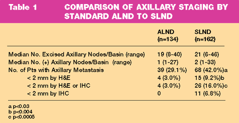
In 1995, axillary
staging in 134 patients with ALND was compared to 162 patients with
SLND followed by completion ALND. One to two sections of all nonsentinel
lymph nodes were evaluated with H&E. Multiple sections of the SLN
were evaluated with H&E and anticytokeratin IHC.
The number of
patients with axillary metastases was 29.1% in the ALND group and
42.0% in the SLND followed by ALND group (p<0.03). The results are
shown in Table 1.
- SLND with
multiple sectioning and IHC staining of the SLN increases accuracy
of axillary staging
- Histopathologic
evaluation of the SLN identifies more patients with axillary metastases
which could be missed on routine H&E examination
- Micrometastases
are easier to identify in the SLN with multiple sections and IHC
|
1994: The Third Challenge Prospective
Validation |
Could the refinement
of SLND and histopathologic evaluation improve the ability to identify
a SLN and accurately stage the axilla over what was originally determined
in the pilot study? By 1994, the experience with ILM and SLND for
breast cancer had grown, the technique had been refined and the
pathologic handling of the SLN was more sophisticated with increased
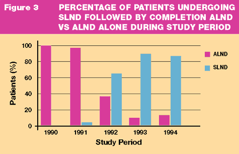
detection of metastases. From the inception of the SLND in 1991,
the number of patients with clinically negative axillae who were
treated with SLND followed by completion ALND increased (Figure
3).
The purpose
of this study was to validate SLND with the mature surgical and
pathologic techniques as accurate and sufficient to stage the axilla
in breast cancer (3).
From July 1994
to October 1995, 107 patients with potentially curable breast cancer
underwent SLND with the mature method. SLND was followed by Level
I, II and part III ALND in conjunction with breast-conserving surgery
or mastectomy. The SLN was evaluated with H&E and IHC. Nonsentinel
nodes were evaluated by H&E.
The median age
of the patients was 56.6 years. The mean tumor size was 2.11 + 1.38
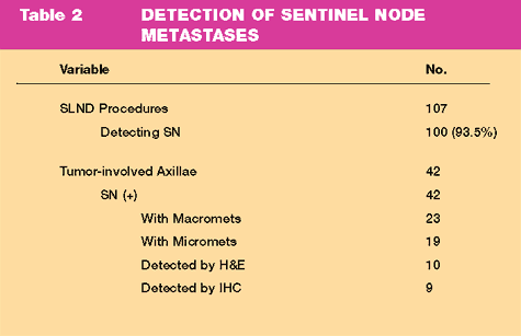
cm. SLN was identified in 100 of 107 patients (93.5%). There were
42 patients with (+) SLN and 28 (66.7%) had no other positive ALN.
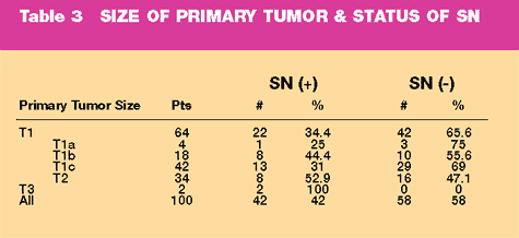
The method of detection is shown in Table 2. The status of the axilla
relative to the tumor size is shown in Table 3. In the seven patients
with no SLN identified, 6 had a tumor free axilla.
- SLND is minimally
invasive
- ILM with
1% isosulfan blue identifies the SLN in 93.5% cases
- SLN histopathologic
analysis with cytokeratin IHC is highly accurate
- SLND was
100% accurate with no false negatives
|
The Fourth Challenge Complete Nonsentinel
Node Staging |
Is the enhanced
detection of metastases, especially micrometastases a reflection
of the intense histopathologic evaluation of the SLN compared to
nonsentinel nodes and does not represent any biologic significance?
The purpose of this study was to determine whether the sentinel
node is truly the axillary lymph node most likely to harbor metastatic
tumor and to assess the true histologic false-negative rate of SLND
at our institution (4) .
From
February 1994 to October 1995, 103 patients underwent SLND followed
by completion ALND. The median age was 55. The median tumor size
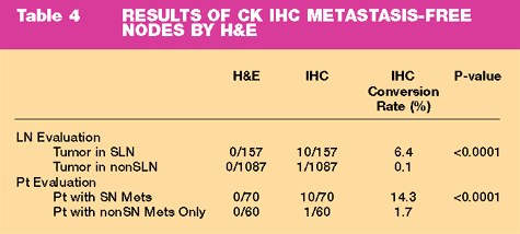
was 1.8 cm. H&E staining identified 33 patients with (+) SLND (32%).
IHC evaluation of 157 (-) SLN upstaged 10 patients (14.3%) (Table
4). In 60 patients whose SLN were negative by H&E and IHC, 1087
nonsentinel nodes were examined at 2 levels by IHC. Only 1 additional
tumor positive node was identified.
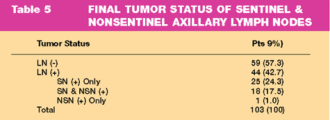
The
final tumor status of sentinel and nonsentinel axillary nodes demonstrates
that SLND alone would be sufficient for 57.3% of patients with no
evidence of axillary metastases. In addition, 24.3% had only involvement
of the SLN (Table 5)
- If the SLN
is tumor free by H&E and IHC, the probability of nonSLN involvement
is <0.1%
- The false
negative rate for nonSLN metastasis is 0.97%
- The SLN is
the most likely node to harbor metastases
|
1995: The Fifth Challenge Assess Feasibility
of A Change In Paradigm |
The
standardization of SLND, the validation with completion ALND and
complete histopathologic assessment of the SLN and nonSLN challenges
the establish paradigm of ALND for staging in breast cancer when
the SLN is negative. In 1995, the ability to identify the SLN was
93.5% and the accuracy of the SLN to predict the status of the axilla
was 99%. The purpose of this study was to determine the complication
rate and the local recurrence rate in women who had a tumor-free
SLN who did not undergo ALND (5).
From October
1995 to July 1997, 133 consecutive women with tumors <4 cm underwent
SLND for staging. SLND was the only axillary operation if the SLN
(-). Completion ALND was performed when the SLN (+).
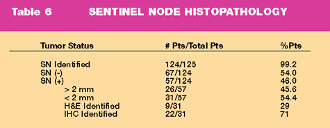
SLND identified
the SLN in 132/133 patients (99% accuracy). Eight patients were
excluded from analysis. Three elderly patients with SLN micrometastases
refused ALND and five because of subsequent ALND. All 5 patients
were SLN (-) and ALND (-). The status of the axilla is shown in
Table 6.
Complications
occurred in 20 patients (35%) undergoing ALND after SLND, but in
only two patients (3%) undergoing SLND alone (p=0.001). There were
no local recurrences at a median follow-up of 39 months.
- The ability
to identify the SLN in breast cancer with intraoperative lymphatic
mapping with 1% isosulfan blue is 99%.
- Minimally
invasive SLND results in statistically significant reduction in
axillary morbidity
- SLND alone
does not demonstrate any adverse outcome for locoregional control
at a median follow-up of 39 months.
|
1999: The Sixth Challenge Multicenter
Validation |
At the close
of the 20th century, the surgical treatment of breast cancer has
turned to refined surgical assessment of locoregional disease to
minimize the morbidity of breast cancer surgery without compromising
locoregional control. The enrollment into multicenter national trials
such as the ACoSOG Z10 and Z11 seek to further delineate the extent
and type of surgical intervention appropriate for the treatment
of breast cancer using ultrastaging. Intraoperative lymphatic mapping
with isosulfan blue and SLND has brought us to a new series of questions
regarding the most appropriate surgical intervention in the axilla,
much like breast conserving surgery did 30 years ago.
Since May 1999,
207 patients at the JWCI have been enrolled in the ACoSOG Z10 Trial.
There was 100% ability to identify the SLN.
| Conclusions
A Decade Of SLND For Breast Cancer |
- A method
was defined
- Improved
Staging was demonstrated with the SLN
- Complete
axillary node assessment was seminal for verification of the SLN
as the first tumor draining node
- Single institution
validation with the refined technique confirmed the principle
- SLND for
node negative patients has become a new paradigm for breast cancer
treatment n SLND is undergoing multicenter validation
- The future
role of SLND in breast cancer is as a fulcrum around which new
clinical trials are designed to answer questions about staging
and multidisciplinary treatment
1. Giuliano,
A., Kirgan, DM, Guenther, JM, Morton, DL (1994) Lymphatic Mapping
and Sentinel Lymphadenectomy for Breast Cancer. Ann Surg, 220, 391-401.
2. Giuliano,
A., Dale, PS, Turner, RR, Morton, DL, Evans, SW, Krasne, DL (1995)
Improved Axillary Staging of Breast Cancer with Sentinel Lymphadenectomy.
Ann Surg, 222, 394-401.
3. Giuliano,
A., Jones, RC, Brennan, M, Statman, R (1997) Sentinel Lymphadenectomy
in Breast Cancer. J Clin Oncology, 15, 2345-2350.
4. Turner, R.,
Ollila, DW, Krasne, DL, Giuliano, AE (1997) Histopathologic Validation
of the Sentinel Lymph Node Hypothesis for Breast Carcinoma. Ann
Surg, 226, 271-278.
5. Giuliano,
A., Haigh, PI, Brennan, MB, Hansen, NM, Kelley, MC, Ye, W, Glass,
EC, Turner, RR (2000) Prospective Observational Study of Sentinel
Lymphadenectomy Without Further Axillary Dissection in Patients
with Sentinel Node-Negative Breast Cancer. J Clin Oncology, 18,
2553-2559.
Top
of Page
|
